Protein Shape Changes Could Reveal Early Disease Signals
Prof. Paola Picotti explains how structural proteomics reveals protein shape changes that help decode disease.
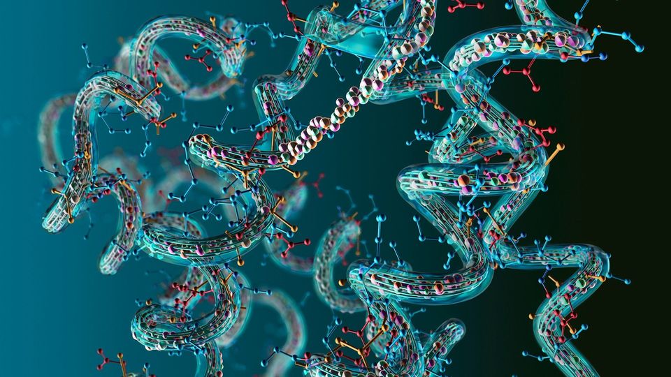
Proteomics is moving beyond measuring protein abundance to understanding how proteins change shape, interact and function within the cell.
Prof. Paola Picotti, a leading figure in proteomics and a full professor in the Department of Biology at ETH Zurich, has been at the forefront of developing innovative mass spectrometry techniques that reveal these dynamic protein landscapes.
At HUPO 2025, Picotti spoke with Technology Networks to discuss the distinctions between classical proteomics and limited proteolysis mass spectrometry, while exploring how protein–metabolite interaction maps can illuminate cellular regulation and guide drug discovery.
Picotti also talked about the potential of structural proteomics to uncover early molecular events in neurodegenerative diseases and shared her perspective on connecting proteome-wide structural data to functional outcomes in disease models.
Could you explain how limited proteolysis mass spectrometry (MS) differs conceptually from conventional bottom-up or native proteomics approaches?
Paola Picotti, PhD (PP):
Classical proteomics, or bottom-up MS, typically tries to measure protein abundances and how they change across conditions. While limited proteolysis MS is a structural proteomics tool that tries to detect proteins that undergo structural alterations across conditions as a readout for functional alterations.
RLS:
How can protein–metabolite interaction maps guide drug discovery or our understanding of cellular regulation?
PP:
In cellular regulation, metabolite protein-binding events can profoundly change the activity of proteins. They can regulate enzymatic activity, for example, acting as competitive inhibitors or allosteric regulators.
Detecting these interactions can help you learn the functional regulation of specific proteins.
In terms of drug discovery, detecting metabolites that bind to new allosteric sites can lead to the development of new molecules that bind to the same sites and regulate, indirectly, enzymatic activity.
RLS:
What has structural proteomics revealed about the early, perhaps reversible, stages of proteome destabilization in neurodegenerative diseases?
PP:
You can use these techniques to probe early events that lead to the formation of protein aggregates in the cell.
For example, we have recently developed a version of the method that can be applied directly inside the cell, and this is very useful to capture so-called “phase separated compartments”. These are not necessarily aggregated forms of proteins; however, some of them can evolve into insoluble aggregates, and so these can be probed within the cell.
Another idea would be to apply the technique to patients with early stages of neurodegenerative diseases to try and detect early molecular events that then evolve into a more serious state, either as a biomarker or to study disease mechanisms.
RLS:
How do you connect proteome-wide structural data with downstream functional consequences, such as altered signaling, metabolism or cell viability, in disease models?
PP:
You can probe exactly those downstream events with the same technique.
Let's assume you have a drug. If you apply the drug to a cell lysate and use this technique, you will most likely detect direct interactions of the drug with the proteins that bind it.
If you apply the drug to intact cells, you will probe a mixture of direct interactions and downstream effects.
So, you can, in fact, use it exactly to identify those pathways that are downstream of target engagement, such as signaling cascades that are activated.
RLS:
As limited proteolysis mass spectrometry becomes more established, how do you envision its integration into clinical or diagnostic workflows?
PP:
In one of our studies, we have shown that protein structures can serve as candidate disease biomarkers. That means that certain protein structures are altered upon disease development, and we focused on Parkinson's disease.
Now, these are only candidate biomarkers, and it's a long way there before they become real biomarkers.
However, should they ever be proven to be usable biomarkers, then the next step would be to turn them into an enzyme-linked immunosorbent assay (ELISA) that measures the aberrant conformations
We would need to generate binders that can be used in such ELISA assay, and for that, we are now developing an approach to derive the 3D coordinates of the pathological and healthy conformations to then possibly design binders through protein design tools.
RLS:
Have any talk sessions or emerging themes stood out to you so far at HUPO 2025 in Toronto?
PP:
I was particularly impressed by the huge number of patient samples that are currently being analyzed with proteomics. I think that's something five to ten years ago we would not have expected.
The field has evolved so much and that we can already process such large cohorts of patient samples.


